|
Welcome! We specialize in custom designed molecular animations (for stand-alone use or integrated into a molecular tour or tutorial) and interactive versions of static molecular figures. Our News and Views (immediately below) is an occasional commentary on visualizing molecules and understanding their functions.
New online 3D figures below!
Molecules in Motion News & Views
Reducing the risk of breast cancer return
June 7, 2008
A recent study suggests that Zometa, a drug that is in use for preventing bone loss during therapy for breast cancer, can also reduce the liklihood of the cancer's recurrence. Zometa is a bisphosphonate: a class of small molecules that have a phosphate attached at each end. The drugs act like poison to osteoclasts, cells that re-absorb bone, by binding to a protein called FPPS (farnesyl pyrophosphate synthase). FPPS is essential to the osteoclasts survival, and when it is blocked by Zometa or other such drugs, the osteoclast cells die, and bone re-absorption is slowed.
View a bisphosphonate in complex with FPPS.
Fatty Acid Synthase
March 4, 2008
I'm teaching this month at UMass Amherst, a month of advanced Molecular Biology. The last time I taught at this level, it was in Biochemistry, and I loved teaching about the function of fatty acid synthase (FAS). FAS is a beautiful, intricate complex of activities. So as I trolled Science for interesting structures to present here, the two structures of FAS that were published last year caught my eye. Check out these mammoth enzymes in FirstGlance in Jmol:
View the "central wheel" of FAS from a fungual organism.
View the "domes" of FAS from a fungual organism.
View the wheel and one dome of FAS from yeast.
Comparing the three structures above should give you a feel for the whole complex, which has a wheel or disc that is centered between two large domed structures. Best to open the first two, then close those windows and open the third - they are huge files!
Read about the structures (requires subscription):
Structure of Fungal Fatty Acid Synthase and Implications for Iterative Substrate Shuttling. Science 316:254 - 261
Structural Basis for Substrate Delivery by Acyl Carrier Protein in the Yeast Fatty Acid Synthase. Science 316:288 - 290.
By the way, if you know the PDB ID code for a structure, you can explore it in 3D instantly at FirstGlance in Jmol: seven different default views, a Find feature, viewing of non-covalent interactions in a few clicks (see "Contacts") and more.
Cool News - Gene Popsicle
August 7,2007
The LA Times today reports:
"Scientists have recovered microorganisms from ancient Antarctic ice and
coaxed it (sic) back to life in the lab, according to a study published today.
The glacial ice acted as a 'gene Popsicle,' preserving DNA that hasn't
circulated in the gene pool for up to 8 million years." Read more...
How cool is that! ;^)
Speaking of DNA, jump in and explore DNA in interactive 3D! This self-guided tour has an introductory, explanatory DNA page as well. These resources are on the fantastic molecular visualization site, MolviZ.Org.
3D Figure in Biochemical Journal
July 27, 2007
Today's issue of the Biochemical Journal features a Molecules in Motion 3D replica figure. The figure, a complex rendering of an EF hand showing the details of coordination bonds and many hydrogen bonds, allows you to rotate the structure to see it from any angle. The figure also features an animation showing where the coordination complex is situated in the EF hand, and has additional controls for re-centering, rocking, and spinning. Great for presentations!
You can explore calcium coordination by the EF hand — in 3D and if you have a subscription to Biochemical Journal, read the review paper it supplements, "Structures and metal-ion-binding properties of the Ca2+-binding helix–loop–helix EF-hand motifs," by Jessica L. Gifford, Michael P. Walsh, and Hans J. Vogel.
If you don't have a subscription to BJ, but want to see some more replica figures, follow this link: More 3D Figures.
The Molecular Divas of Evo-Devo
June 26, 2007
Ever wonder how the first wing, jaw, or beak came to erupt from a wingless, jawless, beakless earth? Yep, me too. C.K. Yoon's article in the NY Times (free, but registration required) lays out the story of a few key genes that when 'tweaked' give rise to some amazingly powerful changes in shape and function during development. This research has required a melding of two fields that were, once upon a time, distinct - evolutionary biology and developmental biology. Hence the name, evo-devo.
Two of the genes that are playing starring roles in the current research are BMP4, a member of the TGFβ family, and calmodulin. Calmodulin's structure has been determined and is well-studied. Explore calmodulin in 3D using FirstGlance in Jmol!
Interactive 3D Figures
Journal Figures Come to Life as 3D Replicas
Biochemical Journal (BJ) is publishing review articles featuring Molecules in Motion interactive figures. BJ websites require a subscription, however, the Soufi & Jayaraman figure below has been made available free. Thanks, BJ!
PRH/Hex: an oligomeric transcription factor and multifunctional regulator of cell fate.
A. Soufi and P.-S. Jayaraman
3D Figure
Paper
Structural biology of plasmid partition: uncovering the molecular mechanisms of DNA segregation. M.A. Schumacher
3D Figure
Paper
The Hsp90 molecular chaperone: an open and shut case for treatment. L.H. Pearl, C. Prodromou and P. Workman
3D Figure
Paper
The histidine phosphatase superfamily: structure and function. D.J. Rigden
3D Figure
Paper
Two independent routes of de novo vitamin B6 biosynthesis: not that different after all. T.B. Fitzpatrick, N. Amrhein, B. Kappes, P. Macheroux, I. Tews and T. Raschle
3D Figure
Paper
Na+/Ca2+ exchangers: three mammalian gene families control Ca2+ transport. J. Lytton
3D Figure
Paper
Structures and metal-ion-binding properties of the Ca2+-binding helix–loop–helix EF-hand motifs. J.L. Gifford, M.P. Walsh and H.J. Vogel
3D Figure
Paper
ACS Chemical Biology papers with Molecules in Motion 3D figures:
HIV-1 Reverse Transcriptase Structure with RNase H Inhibitor Dihydroxy Benzoyl Naphthyl Hydrazone Bound at a Novel Site
3D Figures (free)
Paper (free)
Mechanistic and Structural Basis of Stereospecific Cβ-Hydroxylation in Calcium-Dependent Antibiotic, a Daptomycin-Type Lipopeptide
3D Figures (free)
Paper (requires subscription)
Structural Basis for High-Affinity Peptide Inhibition of Human Pin1
3D
Figures (free)
Paper (requires subscription)
Other Examples of 3D Interactive Figures
Compare still figures and the interactive replicas, side-by-side.
Any molecular structure figure can be adapted for viewing in 3D on a web page. There is no need to install any software for viewing them. They appear in ordinary web pages.
View Example
For more information, view our flyer, or send an emai with your enquiry to friedar@moleculesinmotion.com.
Animations & Tutorials
Looking for fully interactive, online animations? You've found them. Molecules in Motion has been providing animations of molecules in 3D that are scientifically accurate, beautiful, and clear for over 10 years. Our animations are being featured at the web sites of leading textbooks and journals, in films, and on television. Custom design is our specialty. Animations are also available in QuickTime and other video/film compatible formats.
Interacting with our animations is as easy as clicking the mouse. Just click and drag to rotate in 3D! Zoom in or out using your scroll wheel (or on a Mac, SHIFT-drag).
The above animations were created as tutorials in collaboration with Science Technologies to accompany the textbook Concepts in Biochemistry, by Rodney F. Boyer (John Wiley & Sons, publishers).

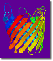
What are Proteins? • View Animation
Composed of twenty different building blocks called amino acids, proteins are the most versatile molecules in biology. Three different proteins illustrate common structural elements of proteins despite their very different functions.
DNA Structure • View Animation
Take a guided tour of DNA in three dimensions. Get to know the double helix from all angles, and the four building blocks that make up the code of life used in DNA. Also includes two movies of RNA. This tutorial was created in collaboration with Science Technologies, to accompany the textbook Essential Biochemistry, by Charlotte W. Pratt and Kathleen Cornely, published by John Wiley & Sons.
Custom Animation Design
Our custom animations are suitable for web delivery, film or video. They have appeared in the National Geographic Channel's Naked Science: "What's Sexy" (we kid you not!) and "Roman Tech", the documentary film, "Flock of Dodos" (about the current evolution and intelligent design controversy), textbooks including Lehninger's Principles of Biochemistry, Stryer's Biochemistry and others. They can be used for supplementing research publications, teaching, presentations, 3D illustration, or any eye-catching purpose you have in mind. For more information, contact us.
Chime Tutorials
For current tutorials, see the Animations section.
Our existing Chime Tutorials are still available below. Chime technology is being replaced by the more versatile Jmol. Our interactive Jmol tutorials are listed above under Animations.
To view our Chime tutorials, you must use Netscape 4.5 - 4.7 and have the Chime plug-in installed. This one-time process then enables you to view hundreds of Chime tutorials, many accessible through the World Index of Molecular Visualization. For more information on Chime, visit MolviZ.Org.
Exploring DNA
An interactive tour for everyone - take a look around a DNA helix! Investigate its nooks and crannies by zooming in and out with the mouse, and turning it this way and that. Click on buttons to watch animations that show off important features of DNA. Includes a version translated to Italian.
DNA & the Double Helix
Designed for classroom presentations, illustrates the fundamentals of nucleotide and double helix structure.
DNA Binding Motifs
In order for the genetic code to be "read" correctly, proteins that attach themselves to DNA are essential. Here are three examples of themes (or "motifs") common to proteins that bind and regulate DNA: the zinc finger, helix-loop-helix, and leucine zipper. Use the buttons to change the colors and representations of the DNA and proteins-- see for yourself how the proteins cling to DNA. Lecture-ready: buttons are labelled with keywords.
Protein Secondary Structure 3.0
The alpha helix and beta sheet are explored in terms of stabilizing forces and interactions within proteins. Includes many hyperlinked definitions of biochemical terms, and questions for discussion. Also available as adapted by Worth Publishing at the Lehninger Biochemistry in 3D web site, where it includes an additional section on beta-turns. (Follow links to "Protein Architechture".) Lecture-ready versions of the alpha-helix and beta sheet are also available.
Protein Secondary Structure 2.0
An overview of the of stabilizing forces, hydrogen bonds, and hydrophobic interactions that characterize protein secondary structures.
Protein Secondary Structure 1.0
For presentation use in a classroom lecture. Movie buttons are labelled with key words instead of textual explanations. The movies allow time for an instructor to narrate.
At the Lehninger's Biochemistry in 3D web site:
G Proteins
Trimeric G Proteins act as molecular switches. In their "on" state, they help relay messages from the extracellular environment to the inside of the cell. Learn how they are activated by the nucleotide GTP.
Hammerhead Ribozyme
All enzymes were thought to be proteins until the first RNA enzyme (ribozyme) was discovered. The Hammerhead is the smallest naturally occuring ribozyme. It appears simple, yet illustrates the salient features of RNA structure and catalysis. Includes an animation using four different conformations of hammerhead, including ground state, two transition states, and products.
Major Histocompatibility (MHC) Molecules
An in-depth look at immune system proteins that enable us to distinguish invaders' molecules from our own. Includes side-by-side comparisons of Class I and II MHC proteins. Also features molecular surfaces for some cool views of the peptide-binding cleft and anchor pockets!
Oxygen Binding Proteins
An in-depth look at two classics: myoglobin and hemoglobin. Includes fundamentals of tertiary structure, in-depth analysis of R to T state conformational switching, the allosteric modulator BPG, sickle-cell disease.
Restriction Enzymes: Eco RV
Restriction enzymes grab DNA and chop it up-- a very useful trick, which gave rise to biotechnology as we know it! Get acquainted with one restriction enzyme, Eco RV, and find out, step by step, how it cuts DNA. Includes an explanation of specific vs. non-specific binding applicable to molecular interactions in general.
Contact Molecules in Motion
Molecules in Motion is owned and operated by Frieda Reichsman, PhD. Feel free to email her for more information about any Molecules in Motion product, or with questions about any of our materials or technology: friedar@moleculesinmotion.com
|